Medical equipment
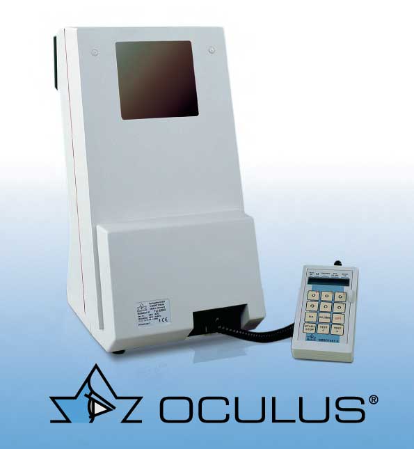
OCULUS MESOTEST II
We are the only refractive laser centre equipped with this device, which is used for more accurate measurement of visual acuity.
MESOTEST II is a device for measuring visual acuity at dusk (in twilight) and sensitivity to glare. The examination uses optotypes of varying contrast, presented against a background of low luminance. The background luminance is 0.032 cd/m² without glare and 0.001 cd/m² with glare. This corresponds to the luminance encountered at dusk or at night. The test optotype is the Landolt ring according to DIN 58220-T1. As visual acuity decreases at low luminance, a visual acuity level of 0.1 is used. Four different contrast levels are available: 1:23 / 1:5 / 1:2.7 / 1:2. Contrast is the ratio of the luminance of the optotype to the luminance of the background. The contrast levels are presented with and without glare. A glare source is built in next to the test field at an angle of 3°; it is activated automatically depending on the selected test. The glare simulates the situation created by an oncoming vehicle with its headlights on full beam. The illuminance of the glare light is 0.35 lux. Mesotest II has three built-in sets of minus lenses, which make it possible to determine so-called night myopia and thus to compensate for it. Mesotest II is operated via an external control unit with a backlit LCD module; it can also be operated from a PC with a software module. Testing visual acuity at dusk and sensitivity to glare is an important addition to daylight visual acuity testing, especially for assessing fitness to drive in the dark. It is also recommended as a pre- and post-operative examination for patients with an IOL (intraocular lens) and for refractive surgery patients, as well as when prescribing tinted spectacle lenses.
PENTACAM®
It helps to describe precisely the condition of the anterior segment of the eye, i.e. the part of the eye from the cornea to the vitreous. It shows corneal thickness in detail, maps the anterior and posterior corneal surfaces and shows the chamber angle, the lens, the ciliary muscle and more. The OCULUS device called Pentacam is a unique combination of precision mechanics, sophisticated software and powerful computing performance. The measurement is fast and contactless!
PENTACAM = 5 important functions:
- Scheimpflug camera
- 3D anterior chamber analyser (e.g. chamber angle, anterior chamber depth, chamber volume, etc.)
- analyser of lens opacity and the extent of cataract
- densitometry, topography of the anterior and posterior corneal surfaces
- full-surface pachymetry (corneal thickness measurement across the entire cornea).

TOMEY® ENDOTHELIAL MICROSCOPE
It can reveal in great detail the condition of the innermost layer of the cornea – the structure and quality of the so-called endothelium. It is the endothelial cells that are responsible for nourishing the cornea and keeping it transparent. The examination is very important before eye surgery, e.g. before cataract surgery, refractive lens exchange, intraocular lens implantation or laser surgery. If the cell count is low or the cells have an unsuitable structure, the doctor will propose a different surgical solution. In this way we prevent complications and ensure your safety. Endothelial microscopy is also used in the diagnosis of congenital corneal disorders, the so-called dystrophies, which usually become apparent only in adulthood or even in old age. The examination is contactless and takes a few minutes. It is completely painless for the patient and places no strain on them. The cornea can also be protected, above all through a healthy lifestyle and by limiting risk factors, which include smoking and spending time in smoky rooms, diabetes, harmful radiation, spending time in air-conditioned rooms and poor air quality.
FUNDUS CAMERA: NIDEK AFC 230 / 210
The AFC-210 non-mydriatic fundus camera has all the functions needed to obtain and evaluate images of the retina quickly and easily. Operator convenience is enhanced above all by automatic focusing on the retina. The device can also capture stereo images and create so-called panoramic images that include the peripheral areas of the retina. It can also take images of the anterior segment of the eye. The high-quality camera captures very high-resolution images, which can be conveniently evaluated and edited with the operating software. The software also includes a patient database in which images can be stored in a clear, organised way and quickly retrieved as needed.
OCT RETINAL TOMOGRAPHY
A retinal scanner with simultaneous OCT and SLO imaging. Further features of the RS-3000 include high image quality achieved with an advanced noise-reduction method, a high scanning speed (53,000 A-scans/s), a 3D image in 1.6 s and ease of use – automatic focusing. Powerful software makes it possible to store measurement results and to analyse captured retinal images in detail in six layers. Follow-up examinations with identical parameters make it possible to monitor and compare the course of pathological changes in the retina.